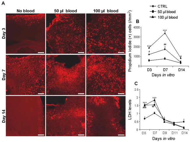
Figure 1. Blood exposure reduces the viability of neural cells in the CS.
A) Representative labeling of cryosections from CS with propidium iodide (PI) at 3, 7 and 14 DIV. Note greater abundance of PI (+) cells in CS treated with blood compared to untreated controls for 3 and 7 DIV; and higher density of PI (+) cells are at d 7 than at d 3 and 14 in blood exposed slices. Scale bar=50 μm. B) Data are mean ± s.e.m (n=4–5 pups). PI (+) cells were significantly more in CS treated with 50 and 100 μl blood compared with controls at d 3 and 7. PI (+) cells were higher in number in CS treated with 100 μl blood compared with 50 μl at 3 and 7 DIV. C) Data are mean ± s.e.m (n=6 pups each group). LDH levels were the highest at day 7 for untreated and blood treated CS. LDH levels were less in untreated CS compared to both 50 and 100 μl blood-treated CS at 3 and 7 DIV. LDH levels were higher in 100 μl blood-treated CS compared to 50 μl treated CS at day 7. *P<0.05, ***P<0.001 for the comparison between 100 μl blood exposure and controls. #P<0.05, ###P< 0.01, ###P<0.001 for the comparison between 50 μl blood exposure and controls.

