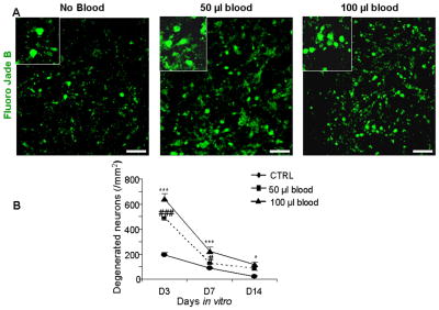
Figure 3. Blood treatment enhances neuronal degeneration.
A) Representative Fluoro-Jade B labeling of fixed cryosections from CS at day 3. Note abundance of degenerating neurons in CS exposed to blood and relatively few in untreated cultured slices. Scale bar, 20 μm. B) Data are mean ± s.e.m (n=5 pups). The density of degenerating neurons was higher in CS treated with 50 or 100 μl blood compared to controls at 3 and 7 DIV. At day 14, degenerating neurons were greater in number in CS exposed to 100 μl blood than controls. ***P<0.001, *P<0.05 for the comparison between 100 μl blood exposure and controls. ###P<0.001, #P<0.01 for the comparison between 50 μl blood exposure and controls.

