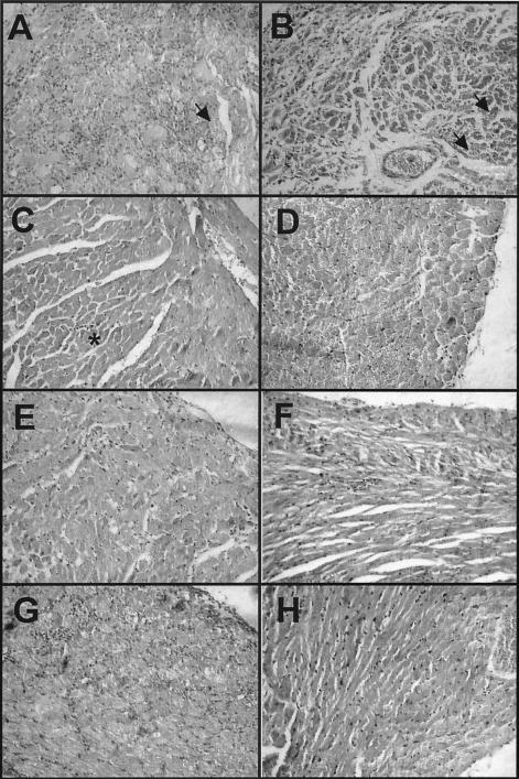
FIG. 2.
Histopathological analysis of cardiac tissue from BALB/c mice infected with a lethal dose of T. cruzi and treated with DNA vaccines. Cardiac tissue was examined on days 40 to 50 postinfection (A, B, C, E, and G) or on day 140 postinfection (D, F, and H). Infected mice treated with a saline solution (A) or the control pcDNA3 plasmid (B) died by days 40 to 45 and had extensive inflammatory infiltrates, scattered to abundant amastigote nests (arrows), and some fibrosis and necrosis. Mice treated with 100 μg of DNA vaccine encoding TSA-1 on days 5 and 12 (C) had only mild focal inflammation (asterisk). When treatment was delayed and administered on days 10 and 17 postinfection (E) or on days 15 and 22 postinfection (G), the inflammation level appeared to be intermediate between that of treated mice and that of untreated mice. On day 140 after infection, BALB/c mice treated with TSA-1 DNA on days 5 and 12 (D) or on days 10 and 17 (F), as well as mice treated with Tc24 DNA (H), all had very mild and diffuse inflammatory infiltrates, and amastigote nests were undetectable except in one mouse.

