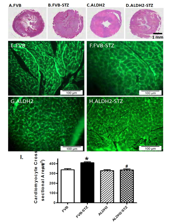
Figure 6.
Histological analyses in hearts from FVB and ALDH2 transgenic mice treated with or without streptozotocin. (A-D) Representative photomicrographs from gross morphological view of transverse myocardial sections (scale bar = 1 mm). (E-H) Representative fluorescein isothiocyanate-conjugated wheat germ agglutinin staining depicting cardiomyocyte size (× 200; scale bar = 100 μm). (I) Quantitative cardiomyocyte cross-sectional (transverse) area from 60 cells from three mice per group. Mean ± SEM, *P < 0.05 versus FVB; #P < 0.05 versus FVB-STZ group.

