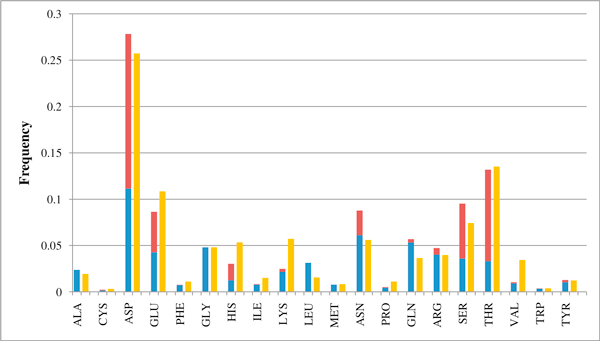
Figure 2.
Frequency distribution of Magnesium binding residues in PDB templates and in annotated human sequences. Distribution of the frequency of residues coordinating magnesium ions in the PDB structures (1,341, blue color: Mg is coordinated by the backbone carbonyl oxygen, red color: Mg is coordinated by the lateral side chain) and in the putatively annotated human sequences (3,751, yellow color).

