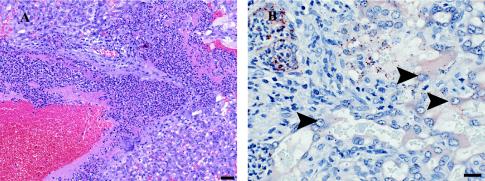
FIG. 2.
Histological examination of guinea pig placental sections. Pregnant guinea pigs between days 42 and 52 of gestation were inoculated intravenously with 2 × 10⁷ wild-type L. monocytogenes bacteria. (A) Hematoxylin-eosin staining of placental section, showing the labyrinthine region at 24 h postinoculation. Moderate numbers of neutrophils and very small numbers of macrophages infiltrate the central region of the lobe. There is fibrin deposition associated within this infiltrate. Bar, 50 μm. (B) Immunohistochemistry for L. monocytogenes reveals large numbers of immunoreactive bacteria within the inflammatory infiltrate. Some bacteria appear to be inside trophoblasts (arrowheads). Bar, 20 μm.

