Figure 3.
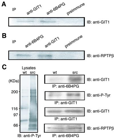
Coimmunoprecipitation of PTPζ and GIT1/Cat-1. (A and B) NIH 3T3 cells were cotransfected with GIT1/Cat-1 and PTPζ-D1902A expression plasmids. Following the transfection, cells were plated on fibronectin. The cell lysates were immunoprecipitated with the appropriate antibodies, and the precipitated proteins were detected with anti-GIT1/Cat-1 (A) and anti-RPTPβ (B) antibodies. (C) Native 3Y1 (wt) and v-src-transformed 3Y1 (src) cells were cotransfected with GIT1/Cat-1 and PTPζ-D1902A expression plasmids. The cell lysates were analyzed by Western blotting using antiphosphotyrosine antibody (Left). The cell lysates were subjected to immunoprecipitation (Right) with anti-6B4 PG (first blot) or anti-GIT1/Cat-1 (second blot) antibodies. Western blotting indicated that GIT1/Cat-1 is highly tyrosine-phosphorylated (second blot) and is present in complex with PTPζ-D1902A (first blot) in the v-src-transformed 3Y1 cells. Both cells expressed comparable amounts of GIT1/Cat-1 and PTPζ-D1902A (third and fourth blots, respectively).

