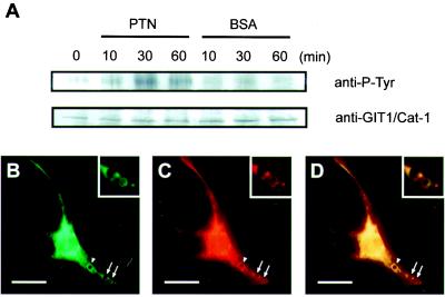
Figure 6.
PTN stimulates tyrosine phosphorylation of GIT1/Cat-1 in the B103 cells. (A) Time course of the tyrosine phosphorylation of GIT1/Cat-1 in response to polystyrene beads coated with PTN (20 μg/ml) or BSA (20 μg/ml). B103 cell lysates (800 μg) were immunoprecipitated with anti-GIT1/Cat-1 antiserum and analyzed by Western blotting with antiphosphotyrosine antibody (Upper) and anti-GIT1/Cat-1 antibody (Lower). PTN-coated beads induced accumulations of GIT1/Cat-1 (B) and PTPζ (C) around the beads after 30 min of incubation (arrows and arrowheads). The combined image (D) shows that the two proteins were coaggregated around the beads. (Insets) A magnified view of the beads marked by arrows. A similar accumulation was also observed around BSA-coated beads (data not shown). [Scale bars, 20 μm (B–D).]

