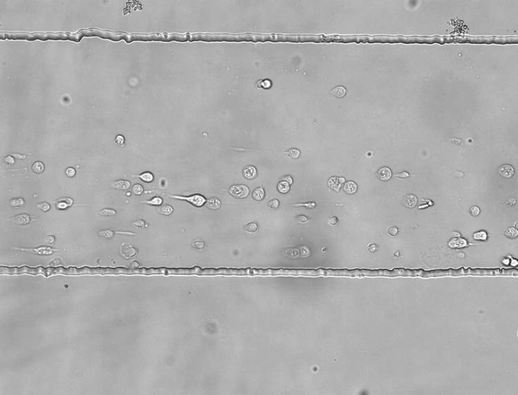
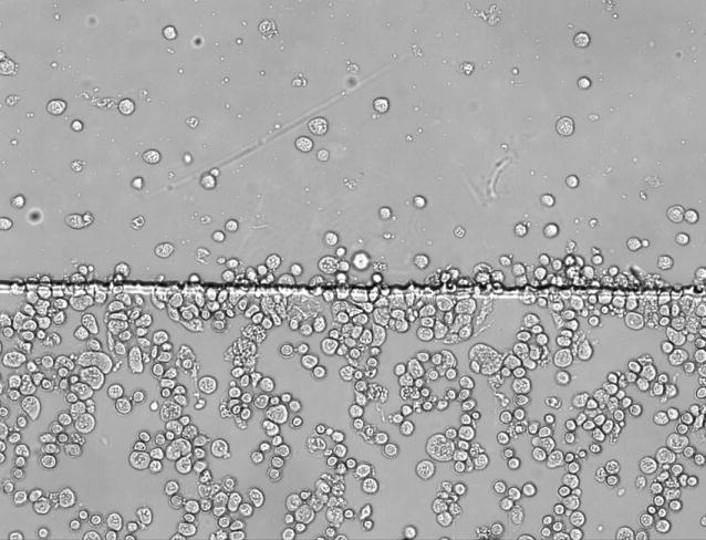
Figure 4.


Cells anchored by anti-CD71 in a single-layer chip (A) and a two-layer reagent delivery chip (B). The reagent channel occupies the upper portion of the figure in each case. In the single-layer chip, the cells are observed to orient in the fluid flow (due to shear stress). In the two layer chip, cell growth is random in direction, indicating minimal influence from shear stress. The flow rate in both cases was 0.1 mL/h for all reagent streams. The scale is the same for both images.
