Figure 5.
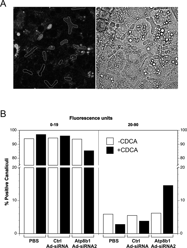
Increased amounts of fluorescent labeled phosphatidylserine (NBD-PS) were observed within bile canaliculi in Atp8b1 knockdown hepatocytes. The cells were treated with PBS, scrambled Ad-siRNA (Ctrl Ad-siRNA), or ATP8B1 Ad-siRNA2 and cultured for 5 days in collagen sandwich gel configuration. Prior to confocal microscopy, the cells were loaded with NBD-PS, and then incubated with or without 5µM CDCA for 16 hr. A. Pseudocanaliculi were first delineated in the phase image and then the outlines were superimposed over the fluorescent image. Shown is one example with positive lumens. B. NBD-PS fluorescence within the outlines was quantitated using Image J. Based on the PBS control levels, canaliculi were divided into 2 groups, below or above the basal fluorescent level of 19. The percentage of canaliculi whose fluorescence intensity fell above or within the PBS control level were then determined for each treatment group. Data represent all canaliculi from 3–4 independent experiments. Bar, 20µm.

