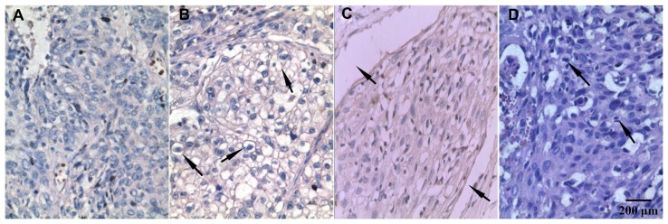
Figure 4.
Immunohistochemistry staining of TRPC6 protein expressed in human cervical carcinoma tissues (100×). (A) There was little to no cell membrane staining when the primary antibody was substituted with phosphate-buffered saline. (B) More than 50% cells stained positive for TRPC6, cell membrane staining was seen at the arrow. (C) TRPC6-stained sample with vascular wall invasion, which was indicated at the arrow. (D) Hematoxylin and eosin staining sample with nuclear atypia at the arrow.
Abbreviation: TRPC6, transient receptor potential channel C6.

