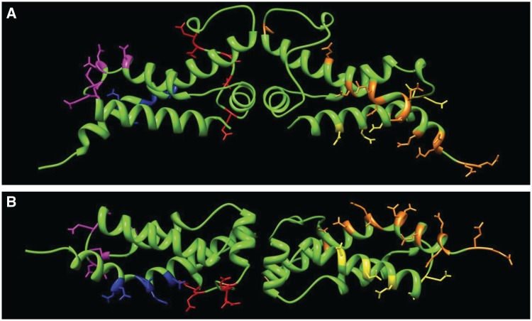
Figure 1.
The location of the Ocr mutants on the dimeric atomic structure (PDB 1S7Z, 4). The residues targeted for mutagenesis are highlighted as coloured sticks. (A) The locations of mutations for Mut2 (magenta), Mut4 (blue) and Mut12 (red) are shown in left-hand monomer and Mut1 (yellow) and Ocr/POcr (orange) in the right-hand monomer. (B) A view rotated 90° from that in panel (A) showing the bottom of the structure.

