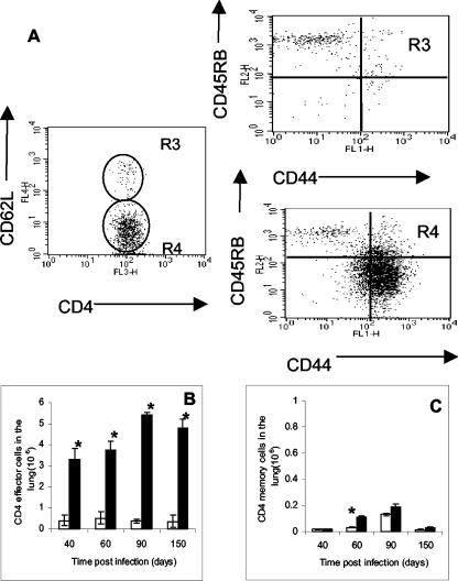
FIG. 2.
Surface marker expression by isolated lung CD4 T cells. (A) In the representative example, lung lymphocytes were stained for CD4 and CD62L cells. CD62Lhigh (R3) and CD62Llow (R4) cells were further analyzed for the expression of CD44 and CD45RB. (B) Kinetics of effector CD4 cells (CD44high, CD45RBmid, and CD62Llow) over the chronic phase of the infection. (C) Kinetics of memory CD4 cells (CD44high, CD45RBlow, and CD62Lhigh). Data are expressed as the means ± the standard errors of the means and are from six mice per group per time point, representative of two independent experiments. Open bars, control aged-matched mice; filled bars, infected mice. Statistical significance based on a comparison of control and infected mice is indicated by an asterisk (P < 0.01).

