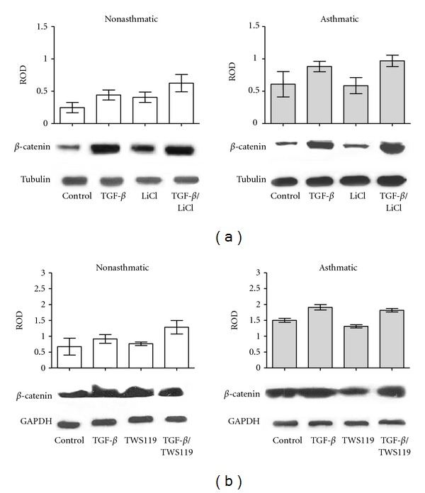
Figure 5.

The effect of inhibition of GSK-3β on β-catenin expression in control and TGF-β 1-stimulated HBFs. HBFs from NA and AS populations were cultured in DMEM supplemented with 0.1% BSA (control) and with the same medium supplemented with TGF-β 1 (5 ng/mL), LiCl (10 mM), or TGF-β 1/LiCl (a) or in DMEM supplemented with 0.1% BSA (control) and with the same medium supplemented with TGF-β 1 (5 ng/mL), TWS119 (10 μM), or TGF-β 1/TWS119 (b) for 7 days. Total protein extracts (30 μg of protein per lane) were subjected to SDS-PAGE electrophoresis, and β-catenin/α-tubulin protein (a) or β-catenin/GAPDH protein (b) was detected by immunoblotting. The graphs in (a) and (b) represent results of densitometric analysis of immunoblots. The amount of β-catenin was normalized to the expression of α-tubulin or GAPDH, respectively. Representative immunoblots from 3 independent experiments are shown under the graphs.
