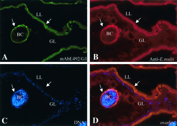
FIG. 1.
Immunofluorescence localization of MAb E492/G1-reactive epitopes in E. multilocularis metacestodes. Sections of LR-White-embedded parasite tissue were stained with MAb E492/G1 and a fluorescein isothiocyanate-conjugated secondary antibody (A), followed by polyclonal rabbit anti-E. multilocularis metacestode antiserum and rhodamine-conjugated goat anti-rabbit IgG (B). The parasite nuclei were stained with Hoechst 22358 to indicate the living parasite tissue (C). (D) Overlay of all three stainings. Arrows indicate the presence of MAb E492/G2-reactivity on the laminated layer (LL) and at the periphery of brood capsules (BC), while the germinal layer (GL) remains unstained.

