FIG. 4.
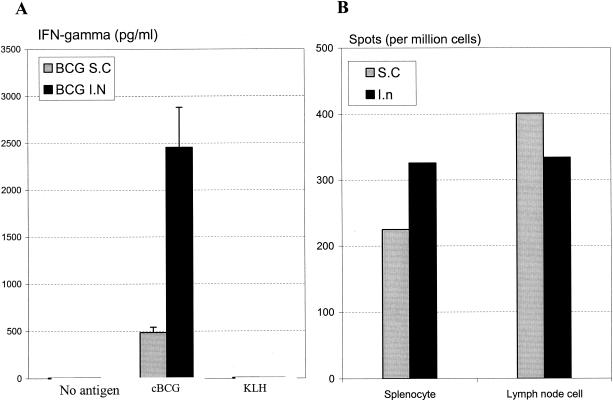
Mycobacterial antigen-stimulated IFN-γ recall release by lymphocytes. BALB/c mice were challenged intratracheally with BCG at 10 weeks after s.c. or i.n. vaccination, and splenocytes from four mice per group were isolated and pooled at 3 weeks postchallenge. Cells were then stimulated with or without mycobacterial antigen cBCG for 72 h. Culture supernatant was assayed for the amount of IFN-γ (A). Results are expressed as means ±standard errors of the means from triplicate determinations. Also, splenocytes isolated at the same time point postchallenge were cultured with mycobacterial antigen M. tuberculosis CFP for 24 h, and the numbers of antigen-specific IFN-γ-producing cells both in spleen and thoracic lymph nodes were determined by ELISpot assay (B). Results are expressed as averages from duplicate determinations.

