
Figure 2.
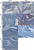
Protein expression of AnxA1 and FPR2 in ankle joints of naive mice. Ankles of naive and K/BxN serum (50+50 μl, day 0 and day 2) were processed (see methods) and stained for AnxA1 and FPR2, with a haematoxylin counterstain. Images representative of three distinct analyses in four mice per status are shown. (A) Naive mouse ankle joints. (B) Serum-treated mouse ankle joint. (C) Same joints but tested without the primary antibody. (D) Joint of an AnxA1−/− mouse acting as negative control for AnxA1 staining. Scale bars, 20 μm.