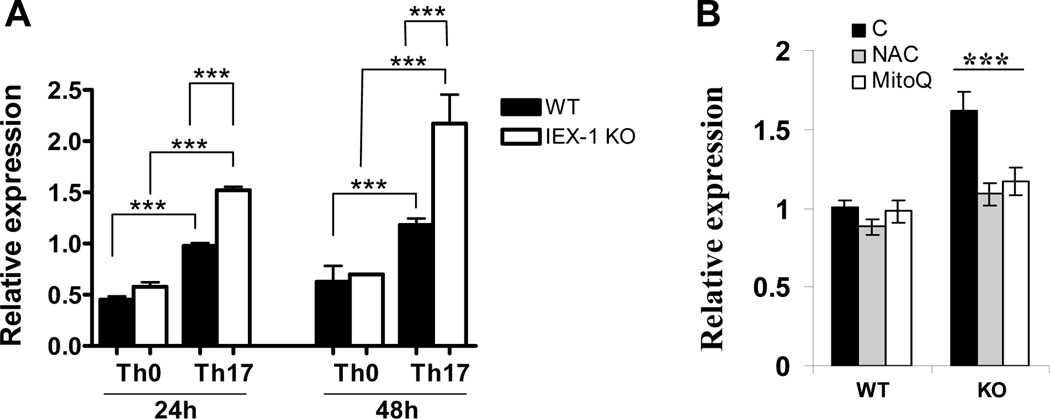
Figure 5.
Increased expression of Batf in the absence of IEX-1 in a ROS-dependent fashion. WT and IEX-1 KO CD4+ T cells were polarized for 24 or 48 hr in Th0 as figure 1 or Th17 conditions as Figure 2 (A). The cells were also differentiated under a Th17-polarizing condition in the presence or absence (B) of either 5 mM NAC or 200 nM MitoQ for 24 hr. Total RNA was extracted from the differentiated cells and Batf expression was measured by quantitative RT-PCR and normalized to the expression level of β-actin. The data are the mean values ± SD of three independent experiments each performed in duplicate. ** and ***, p<0.01 or 0.001, respectively, in presence or absence of IEX-1 or between Th0 and Th17 cells for A or in the presence or absence of indicated oxidants (B).

