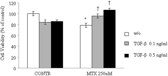
Figure 2. Effect of TGF-β and MTX on cell viability of Caco-2 cells (Alamar Blue reduction test).

Cells were cultured in 96-well plates at a density of 4×10⁴ cells per well up to asubconfluent monolayer (80%), and then they were treated with 0.1 and 0.5 ng/ml TGF-β or control medium for 48 h,and then treated with 0.1 and 0.5 ng/ml TGF-β or MTX 125 nM, or 0.1 ng/ml and 0.5 ng/ml TGF-β with MTX 125 nM or control medium for 72 h. After the treatment, 20% of Alamar Blue was added to each well, and cells were incubated at 37°C for 3 h. Optical density was measured spectrophotometrically at 570 and 630 nm. Cell viability was calculated as percentage of the difference between the reductions of Alamar Blue in treated versus control. Results are presented as percentage of controls, mean ± SEM. CONTR-control, MTX- methotrexate, TGF-β- transforming growth factor beta. *P<0.05 MTX versus control rats, †P<0.05 MTX-TGF-β 0.1 ng/ml and MTX-TGF-β 0.5 ng/ml versus MTX rats.
