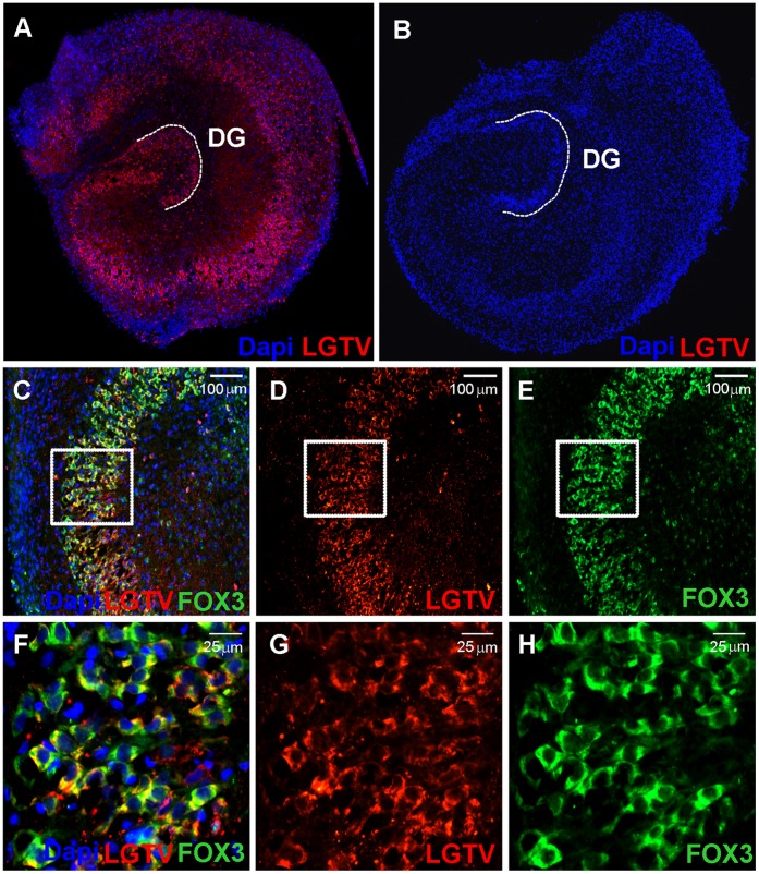
Figure 4. Expression of Langat virus proteins in infected organotypic hippocampal cultures.
OHCs were infected with 2×10E6 FFU Langat virus for 7 days and immunostained with an anti-Langat virus antibody (LGTV; red) (A). Uninfected slices are shown as a control (B). Double staining of viral proteins (LGTV; red) (D 10×; G 40×) and neurons (FOX3; green) (E 10×; H 40×) on infected OHCs showed colocalisation (C 10×; F 40×). Cell nuclei were counterstained with Dapi (blue).

