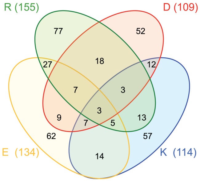
Figure 1. Venn diagram of TM segments (the central 19 residues) containing charged residues: Asp (D, red), Lys (K, blue), Glu (E, yellow) and Arg (R, green).

The value in parenthesis is the total TM helices that contain at least one of such residues. The values inside the ellipses indicate the number of TM helices in each combination of these four amino acids. For example, there are 57 TM helices with only Lys as a charged residue, 12 helices with only Lys and Asp, 7 helices with Lys, Asp and Glu, and 3 helices with all four ionizable residues.
