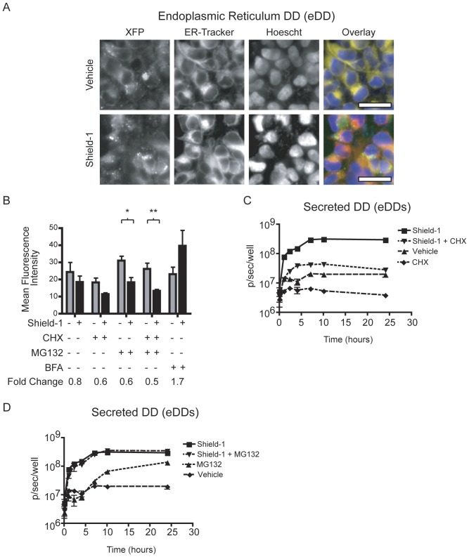
Figure 3. ER and secreted destabilizing domains.
(A) Fluorescence micrographs of eDD cells. The overlay image shows eDD (green), ER-Tracker Red (red), and Hoechst stain (blue). (B) Flow cytometry of eDD cells with Shield-1 (2 µM) or vehicle control after a 6 hour incubation. Cells were co-treated with cycloheximide (CHX, 5 µg/mL), MG132 (5 µM), or brefeldin-A (BFA, 2.5 µg/mL). * P-value<0.05, ** P-value<0.005. (C) Bioluminescence quantification of media from eDDs cells after exposure to vehicle control, Shield-1 (1 µM), CHX (1 µg/mL), or co-treatment with both Shield-1 and CHX. (D) Bioluminescence quantification of media from eDDs cells after exposure to vehicle control, Shield-1 (1 µM), MG132 (1 µM), or co-treatment with both. Error bars represent ± S.E.M. (n = 3).

