
Figure 2.
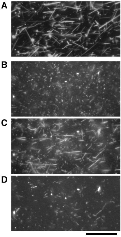
Binding of the TMR-labeled thin filaments to the thick filaments observed with dark-field and fluorescence microscopy. (A) Thick filaments observed in dark-field illumination. (B–D) Thin filaments observed in fluorescence illumination. When the thin filaments were applied onto thick filaments pretreated with cAMP and then washed, they did not remain bound to the thick filaments (B). After the thick filaments were treated with soluble fraction in the presence of ≈10−4 M free Ca2+, thin filaments bound to the thick filaments at ≈10−8 M free Ca2+ (C). Addition of the soluble fraction in the presence of cAMP caused detachment of these thin filaments from the thick filaments (D). All photographs were taken with the same field of view. (Bar = 20 μm.)