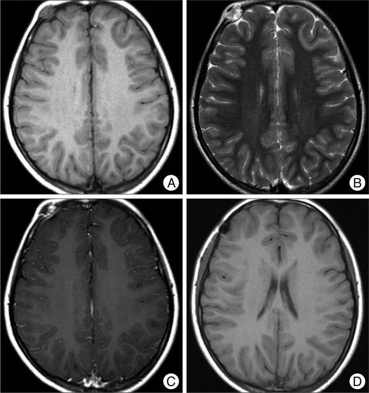
Fig. 2.
Magnetic resonance (MR) image discloses the presence of a 1.3×1.2×1 cm nodular mass skull lesion. This mass lesion is hypointense on T1 (A) and inhomogenously hyperintense on T2-weighted MR image (B) and show inhomogenous enhancement of the soft tissue filling the punch-out lesion (C). Postoperative 1-month T1-weighted axial MR image showing no residual mass with frontal surgical defect (D).

