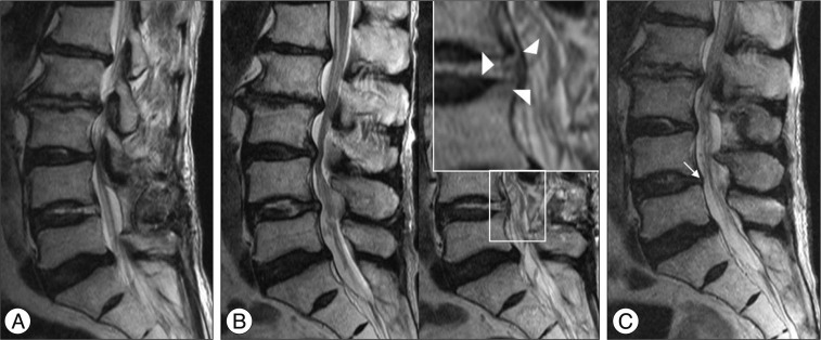
Fig. 2.
A : After discectomy at the L4-5 level, magnetic resonance image (MRI) shows complete removal of the herniated disc. Note the showed cerebrospinal fluid signal in the L4-5 disc space, which is suggestive of dural tear. B : MRI taken when the patient complained of newly developed sciatic pain after discharge. Left sagittal MRI shows no significant interval change from the immediate postoperative MRI (A). However, a nerve root had herniated (arrowheads) into the intervertebral disc space, as shown by the gull wing shape. C : MRI after additional decompression at the left L3-4 and L5-S1 levels. Note the gull wing-shaped nerve root herniation (arrow), which contacts the intervertebral disc.

