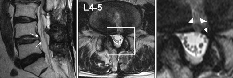
Fig. 3.
MRI showing nerve root herniation (arrows) into the disc space. The point where the nerve root meets with the intervertebral disc space shows a darker signal than that in Fig. 2C. The nerve root formed a loop inside the annulus (arrowheads) on the T2-weighted axial scan.

