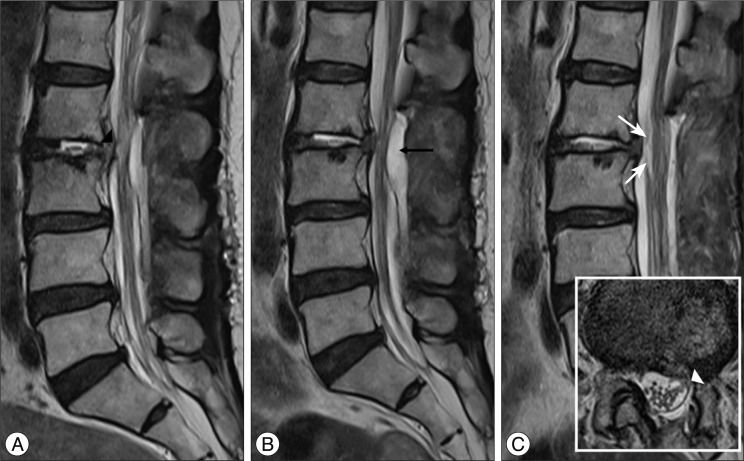
Fig. 6.
Magnetic resonance image performed after additional decompression at the L3-4 and L4-5 levels. The attachment of the nerve root at the L2-3 disc space (black arrowhead) and showed cerebrospinal fluid signal in the L2-3 disc space, which are suggestive of dural tear, can be seen. B : Follow-up MRI showing an epidural hematoma at the L2-3 level (black arrow), which was removed by computed tomography-guided needle aspiration. C : MRI after aspiration of the epidural hematoma. The attachment of the ventral nerve root at the L2-3 disc space is seen in the shape of a gull wing (white arrows) on the sagittal scan, and the attachment of the rootlets on the ventral dura can be seen on the axial scan (white arrowhead).

