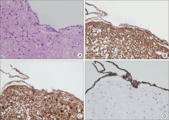
Fig. 3.
In the H&E staining (A), the cyst wall consists of glial cells lined by a simple cuboidal to columnar epithelium. An immunohistochemical examination of the cells lining the cyst wall is positive for glial fibrillary acidic protein (B), S-100 protein (C), and cytokeratin (D). These findings are consistent with an ependymal cyst diagnosis.

