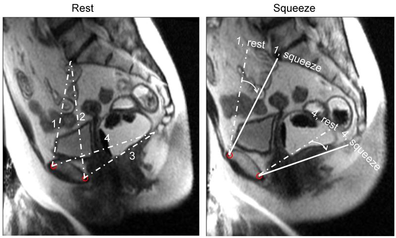
Figure 5. Assessment of pelvic rotation by MRI.
Left panel shows anteroposterior conjugate (1), obstetric conjugate (2), anteroposterior outlet (3), and pubococcygeal line (4) at rest. During squeeze (right panel), observe pelvic rotation as measured by the angle between anteroposterior conjugate and anteroposterior outlet diameters at rest (dashed line) and (solid line) in a control subject.

