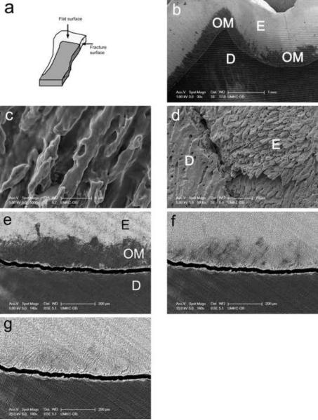
Fig. 1. Identification of interphase organic matrix layer within enamel of mature teeth near the dentin enamel junction.
a, a diagram illustrating shape and orientation of tooth sections used in this study.
b, specimen processed according to “minimalist” protocol and observed without coating at 1 kV accelerating voltage.
c, the microstructure of organic matrix of the “minimalist” specimen, observed on Au-Pd coated specimen.
d, the microstructure of underlying enamel rods after removing of organic matrix from “minimalist” specimen with sodium hypochlorite.
e, specimen processed according to “minimalist” protocol and backscattered electron image collected at 5 kV.
f, specimen processed according to “minimalist” protocol and backscattered electron image collected at 15 kV.
g, specimen processed according to “minimalist” protocol and backscattered electron image collected at 25 kV.
Individual bars refer to microscopic scales for each image.

