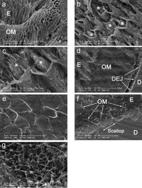
Fig. 3. Identification of sheath-like structures within the enamel interphase matrix.
a, appearance of organic enamel matrix at flat surface of tooth section after etching in EDTA for 90 min, fixation in glutaraldehyde, post fixing in OsO4 and critical point drying.
b, interphase matrix at fracture surface after prefixation for 3 days, etching in phosphoric acid for 15 sec, fixation in glutaraldehyde, post fixation in OsO4, and critical point drying. Asterisks refer to mineral crystals within sheath-like structures.
c, higher magnification view of same specimen as in B. Asterisks refer to mineral crystals within sheath-like structures.
d, appearance of region near the dentin-enamel junction at flat surface after prefixation for 6 days, etching in EDTA for 60 min, fixation in glutaraldehyde, and dehydration with HMDS.
e, higher magnification view of same specimen as in D.
f, appearance of region at fracture surface after prefixation for 6 days, etching in EDTA for 30 min, fixation in glutaraldehyde, post fixation in osmium tetroxide and critical point drying with built in magnetic stirrer turned on (jagged edge of dentin-enamel junction readily visible, arrows).
g, higher magnification view of same specimen as in F (collagen bundles readily visible).
Individual bars refer to microscopic scales for each image.
KEY: E, enamel; OM, organic matrix; D, dentin; DEJ, dentin-enamel junction

