
Figure 3. Combined therapy with Treg and rapamycin inhibits CD4+ and CD8+ T-cell proliferation and potentiates apoptosis of CD4+ lymphocytes in vitro.
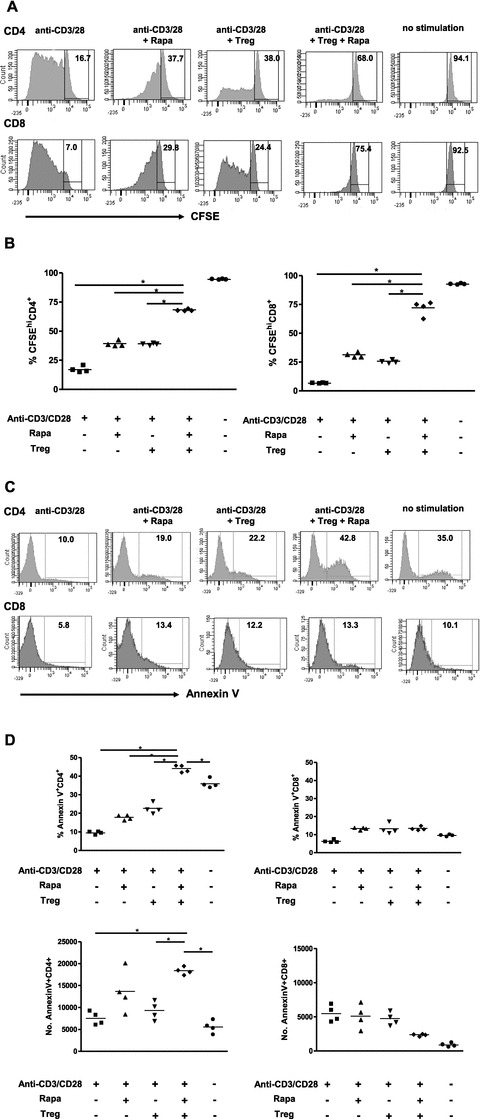
CFSE-labeled PBMC (105 per well) have been incubated with anti-CD3/anti-CD28 beads (cells:bead ratio 5:1) in the presence or absence of 10 nM rapamycin and/or 104 ex vivo expanded CD127lo Treg cells per well. (A) Representative plots depicting CFSE dilution in CD4+ and CD8+ lymphocytes after 5 day of culture. The numbers represent percentage of undivided CFSEhi cells in the gated populations. (B) Percentage of undivided CFSEhi lymphocytes of the total lymphocyte population, as shown in A. (C) Representative plots demonstrating the binding of the apoptosis marker Annexin V to CD4+ and CD8+ T cells. The numbers represent percentage of AnnexinV+ cells of the total lymphocyte population. (D) Absolute numbers and percentage of AnnexinV+ apoptotic cells within the CD4+ and CD8+ gates as demonstrated in panel C.