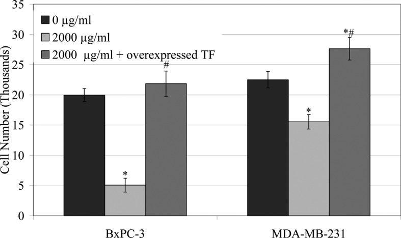
Figure 2.

Influence of LMWH on cellular invasion in BxPC-3 and MDAMB-231 cells overexpressing TF. BxPC-3 and MDA-MB-231 cells were transfected with the pCMV-XL5-TF plasmid and permitted to overexpress TF for 48 h prior to assaying. Cells (105) were placed in the upper chambers of collagen IV-coated Boyden chambers and made up to 250 μl with media containing LMWH (2,000 μg/ml). Media (250 μl) containing 5 μg/ml of bFGF were placed in the lower chambers and incubated at 37°C for 24 h. The number of invading cells was then analysed as in Fig. 1 (*p<0.05 vs. respective untransfected/untreated sample; #p<0.05 vs. respective untransfected/LMWH treated sample).
