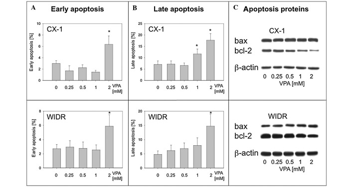
Figure 5.
The presence of apoptosis was analyzed in the cell cultures after a 5-day period of valproate (VPA) exposure with the Annexin V FACS assay. The percentage of (A) early and (B) late apoptotic signs for CX-1 (upper panel) and WIDR (lower panel) cultures is shown. The proportion of cells undergoing apoptotic alterations increased significantly under VPA treatment. The maximum response was found with 2 mM VPA when compared to the untreated controls. (C) From the cell cultures, the corresponding protein extracts were also analyzed for apoptosis-related proteins (bax and bcl-2). β-actin served as an internal control.

