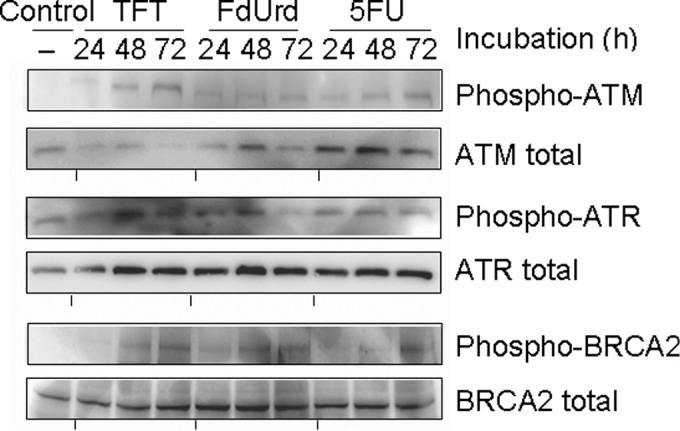
Figure 2.

Western blot analysis of the phosphorylation levels for ATM, ATR and BRCA3 after 24, 48 or 72 h of exposure to the IC50 concentrations of TFT, FdUrd or 5FU in HeLa cell nuclear extracts. The blots are representative of three independent experiments.
