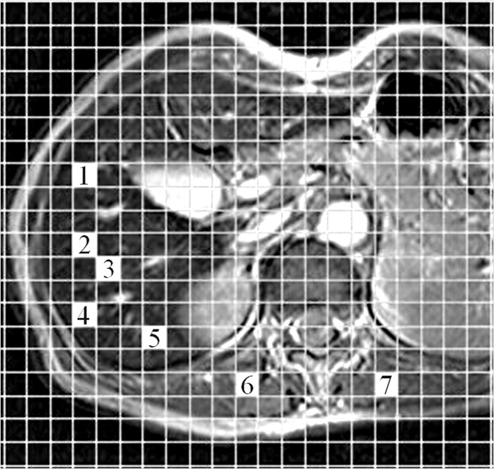
Figure 1.
Measurement method of regions of interest (ROIs) on MRI. Using computer with the plug-in software, two independent observers freely and easily selected an ROI by clicking a mesh unit on an MR image for avoiding the large vessels, focal hepatic lesions or artifacts. One area shown on the MR image is 100 mm2. For liver parenchyma, 5 areas (1–5) were chosen and two areas (6 and 7) were chosen for paraspinous muscle.

