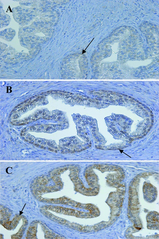
Figure 1.

Immunohistochemical localization of RARα, β and γ in normal human prostate. Immunohistochemical staining was carried out on paraffin-embedded sections (4-μm thick), using primary antibodies as follows: (A) anti-RARα (1:100 dilution), (B) anti-RARβ (1:100 dilution) and (C) anti-RARγ (1:150 dilution). The Envision system was used as the secondary antibody. Original magnification, x200.
