Figure 4.
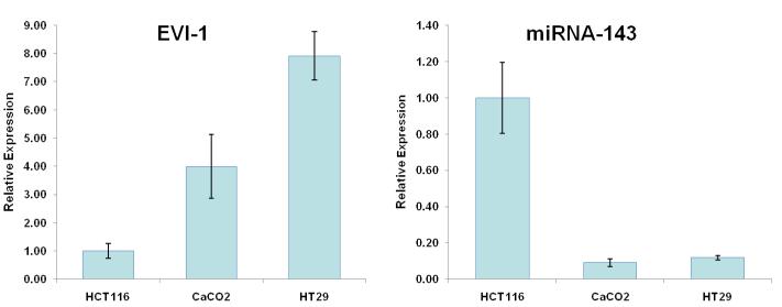
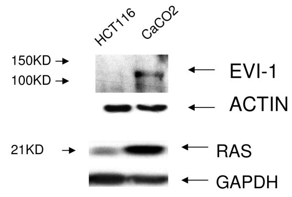
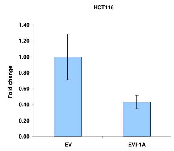
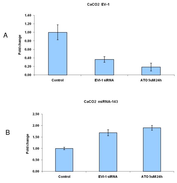
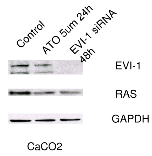
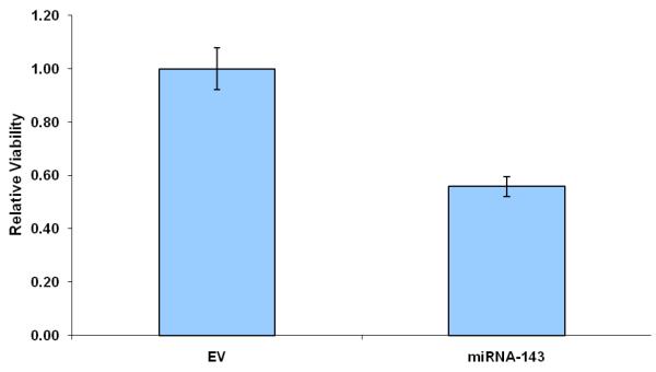
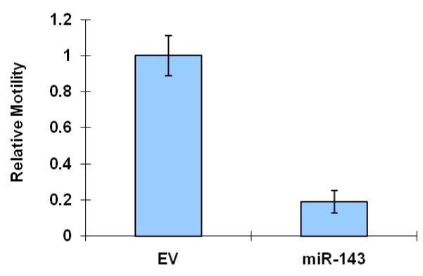
We profiled levels of pri-miRNA-143 and Evi1 in three colon cancer cell lines (CaCO2, HT29 and HCT116). (a) We observed an inverse relationship between levels of Evi1 and pri-miRNA-143, as measured by Real Time PCR. (b). Western blotting revealed a direct relationship between levels of Evi1 and K-Ras in HCT116 and CaCO2 cells. Beta actin and GAPDH were used to normalize RNA and protein input for RT-PCR and western blot, respectively. (c). Overexpression of Evi1 in HCT116 cells by transient transfection of an expression plasmid led to a ~50% reduction in pri-miRNA-143 levels. In contrast, depletion of Evi1 in CaCO2 cells by specific siRNA or arsenic trioxide (ATO) both for 24h led to a ~2-fold increase in pri-miRNA-143 levels (d). Western blot of cells treated with either anti-Evi1 siRNA (48h) or ATO (24h) revealed a decrease in levels of Evi1 and K-Ras proteins in CaCO2 cells (e). We measured the effect of miRNA-143 over expression in Caco2 cells by MTT assay (f) and motility assay (g). Over expression was associated with decreased proliferation and motility, respectively.

