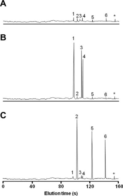
Fig. 3.

Electropherograms of the chemokine profiles of the three subject groups. (A) Normal subjects (Group 3); (B) Patients with neutrophil infiltrations (Group 1); (C) Patients with monocyte and lymphocyte infiltrations (Group 2). Chemokines eluted in the following order: 1. CXCL8, 2. CCL5, 3. CXCL1, 4. CXCL5, 5. CCL3, 6. CCL1, *free dye.
