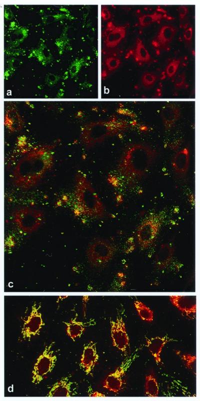
Figure 1.

Colocalization of the α- and β-subunits of ATP synthase on the surface of HUVECs by immunostaining and confocal microscopy. (a) Nonpermeabilized HUVECs immunostained with a murine mAb specific for the α-subunit of ATP synthase. (b) The same cells immunostained with a rabbit polyclonal antiserum specific for the β-subunit of ATP synthase. (c) Composite colocalization images obtained by digital overlays of the above images. (d) Colocalization image obtained from cells permeabilized with ethanol (100%). Representative images are shown; n = 26.
