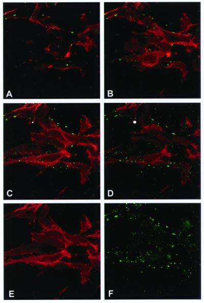
Figure 2.
Surface localization of the α-subunits of ATP synthase and CD31 on nonpermeabilized HUVECs by immunostaining and confocal microscopy. (A–D) Confocal optical sections were taken along the z axis every 1.5 μm. Each section is ≈0.6 μm in thickness. A series of z sections from a representative field is shown, starting with the basal surface in A and ending with the apical surface in D. (E) The same section shown in C; fluorescence from red channel only. (F) The same section shown in C; fluorescence from green channel only; n = 3.

