Figure 10.

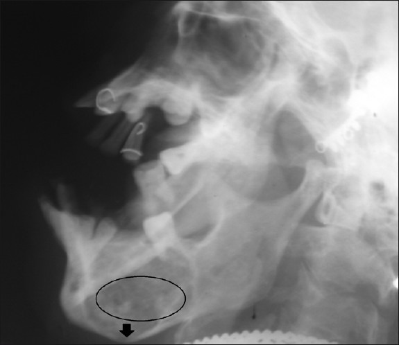
Right lateral skull radiograph shows mixed radiolucent and radiopaque lesion (circle) surrounded by a corticated border in relation to posterior mandible. There is thinning and bowing of the inferior border of the mandible (black arrow).

Right lateral skull radiograph shows mixed radiolucent and radiopaque lesion (circle) surrounded by a corticated border in relation to posterior mandible. There is thinning and bowing of the inferior border of the mandible (black arrow).