Figure 3.

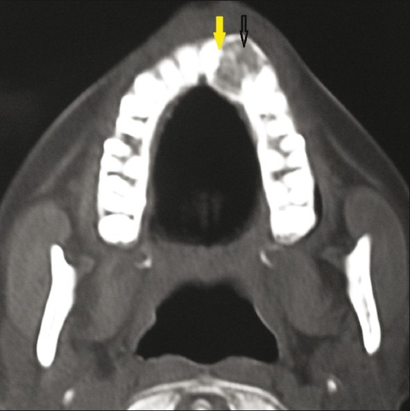
CT Para nasal view shows an expansile lesion with well-demarcated margins and area of calcification (black arrow) in the left maxilla with a displaced maxillary left lateral incisor (yellow arrow).

CT Para nasal view shows an expansile lesion with well-demarcated margins and area of calcification (black arrow) in the left maxilla with a displaced maxillary left lateral incisor (yellow arrow).