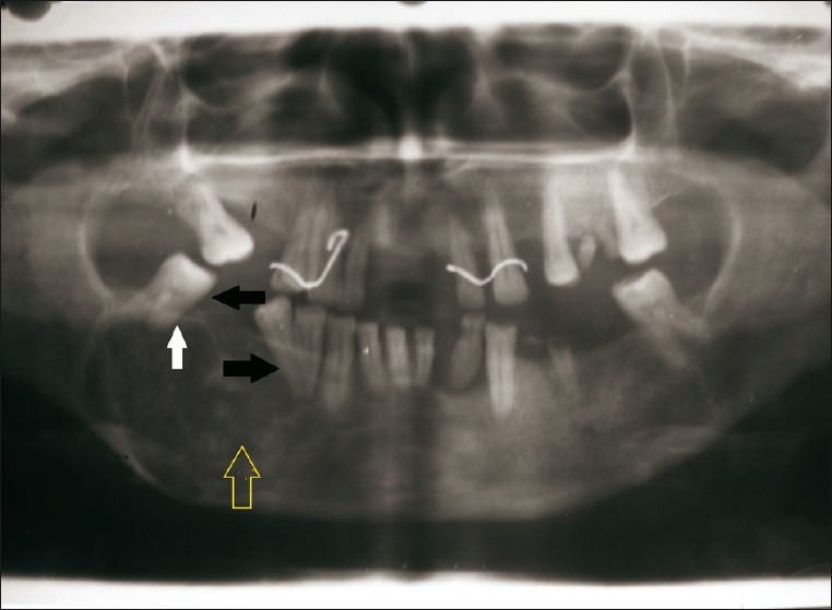
Figure 9.

Orthopantograph reveals a mixed radiolucent-radiopaque lesion (yellow arrow) extending from mandibular first premolar to molar region. The image shows displacement of mandibular right second molar and second premolar (black arrow). There is evidence of root resorption in mandibular right second molar (white arrow).
