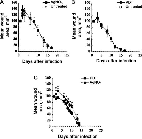
Figure 6.
Mean areas of wounds from mice (n = 6/group) bearing 2 wounds/mouse. Wounds were measured daily, in 2 dimensions, and areas were calculated. Bars indicate SD; *2-tailed P < .05 (unpaired Student's t test). A, Neither wound infected. In each mouse, 1 wound (3 left and 3 right) was treated with photodynamic therapy (PDT) (50 μL of 200 μM ce6 equivalent poly-L-lysine–ce6 solution followed after 30 min by 240 J/cm2 red light). B, Neither wound infected. In each mouse, 1 wound (3 left and 3 right) was treated with 50 μL of 0.5% solution of AgNO3. C, Both wounds infected. In each mouse, 1 wound was treated with AgNO3 and the other with PDT, as described above (3 left and 3 right).

