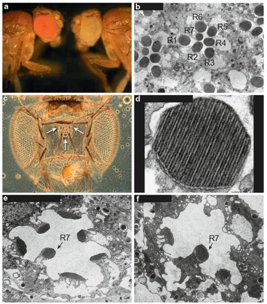
Fig. 52.1.
(a) Wild-type red-eyed fly, Canton S compared to a white-eyed mutant fly, w1118. (b) Cross section through the compound eye showing the R1-7 photoreceptor cells and their photosensitive rhabdomeres (R). The R8 photoreceptor cell is located below the plane of the section. (c) The adult Drosophila visual system showing the two compound eyes and the three simple eyes (ocelli) located on the top of the head (arrows). (d) A higher magnification of a rhabdomere showing the microvilli. The rhabdomeres are made up of about 60,000 microvilli and are 50 nm in diameter and 1-2 mm in length. (e) A newly eclosed ninaEI17 mutant fly, showing the reduced size of the rhabdomeres. ninaEI17 is a null allele, so the flies completely lack Rh1 rhodopsin expressed in the R1-6 photoreceptor cells. (f) Six-day-old ninaEI17 fly, showing that the rhabdomeres of the R1-6 photoreceptor cells are almost completely gone, but the R7 cell rhabdomere remains

