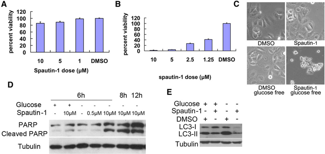
Figure 2. The Biological Effects of Spautin-1 on Cellular Models of Cell Death.
Bcap-37 cells were treated with indicated compounds in normal DMEM with 10% bovine serum (A), glucose free condition (B) or both (C-E) for 48 hr. The cell viability was determined by MTT assay (A), (B), imaged using a phase contrast microscope (C) or the cell lysates were analyzed by western blotting using anti-PARP (D), anti-LC3 and anti-β-tubulin (as a control) (E). All error bars indicate STD. See also Figure S3.

