
Figure 7.
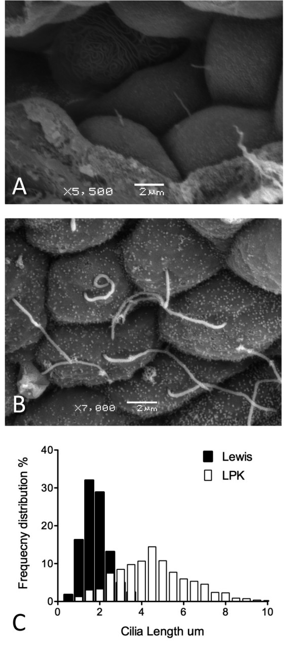
Determination of cilia structure in the LPK. Scanning electron micrographs of renal epithelial cells from Lewis control ( A) and LPK ( B) kidney. The median length of primary cilia is greater in the LPK and they show a greater range of cilia lengths ( C). Scale bars in panels A & B are indicative for that panel.