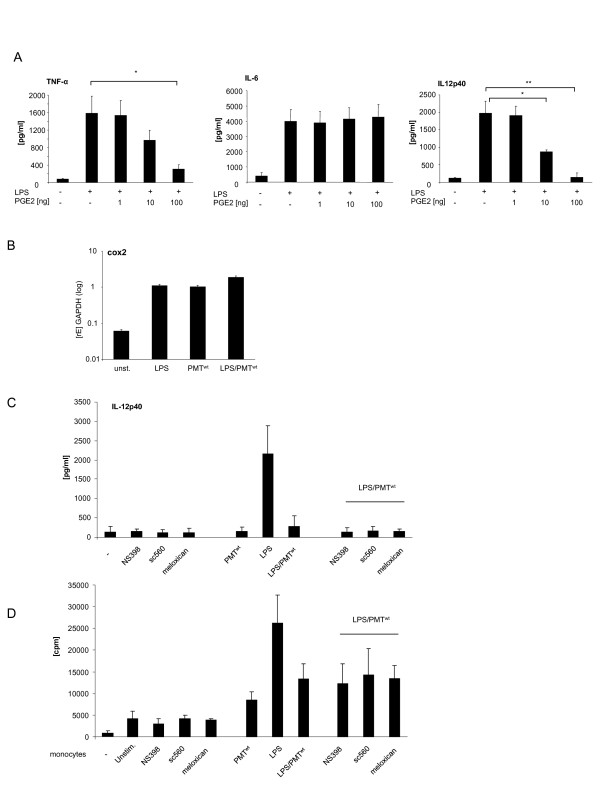
Figure 2.
Contribution of the COX-2/PGE2 pathway to the PMT-mediated modulation of LPS-activated monocytes. A. Monocytes were stimulated overnight with LPS (30 ng/ml) and treated in parallel with PGE2 in increasing concentrations. Supernatants were used to quantify the release of TNF-α, IL-6 and IL-12p40 by ELISA. Presented is the mean and standard deviation of three independent experiments. B. Cells were stimulated with LPS (30 ng/ml; 1 h), PMTwt (1 μg/ml; 4 h) or PMTwt/LPS (4 h/1 h) or left unstimulated. RNA was extracted, cDNA was prepared and used for quantitative PCR analysis with specific primers for COX2. The results are presented as relative expression to the reference gene (GAPDH). Shown is one representative experiment with 10% standard error. C. Monocytes were pre-treated for 2 hours with the cox-inhibitors NS398 (3.8 μM), sc560 (6.3 μM) or meloxican (4.7 μM). Then cells were stimulated with PMTwt (1 μg/ml, 3 h prestimulation) and/or LPS (30 ng/ml; over night stimulation). The release of IL-12p40 was measured by ELISA. Shown is the mean of three independent experiments (mean ± SD; n = 3) D. Monocytes were stimulated as described in C. After washing, the cells were used for MLR with 1x10⁴ CD14+ hBDMs and 1x10⁵ CD3+ alloreactive T cells. After three days of co-culture, proliferation of T cells was analysed by [3H]-thymidine incorporation and presented as counts per minute (cpm). Statistical significance was calculated by using student’s t test with *P < 0.05 and **P < 0.01.

