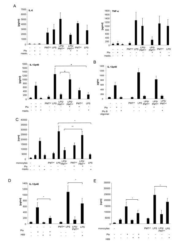
Figure 3.
Gαi-mediated modulation of LPS-induced IL-12 production. A. Monocytes were stimulated overnight with Pertussis toxin (Ptx; 200 ng/ml) or left unstimulated. The next day PMTwt (1 μg/ml) or mastoparan (20 μM) was added. After three hours LPS was added for overnight incubation (30 ng/ml). The release of TNF-α, IL-6 and IL-12p40 were measured by ELISA. Shown is the mean of three independent experiments (mean ± SD; n = 3). B.As additional control cells were treated with Ptx B oligomer and ELISAs were performed with collected supernatant. (mean ± SD; n = 2) C. Monocytes were stimulated as described in A. After washing, co-cultures of CD14+ hBDMs and CD3+ alloreactive T cells were prepared. After 3 days proliferation of T cells was analysed by [3H]-thymidine incorporation and presented as counts per minute (cpm), mean ± SD; n = 3. D. Cells were stimulated as followed: H89 (10 μM; 2 h pre-stimulation) and LPS (30 ng/ml) or Ptx (200 ng/ml) overnight, PMTwt (1 μg/ml, 3 h pre-stimulation) and LPS overnight. Supernatants of cells were used for IL-12 ELISAs. The results are presented as mean ± SD; n = 3. E. Equally treated monocytes were used for MLRs with alloreactive T cells. Shown is the mean and standard deviation of three independent experiments. Statistical significance was assessed by using student’s t test with *P < 0.05 and **P < 0.01.

